Etiology: Clostridium piliforme is a Gram-negative, obligate intracellular, spore-forming rod.
Incidence: The incidence of infection with Clostridium piliforme is rare.
Transmission: C. piliforme is transmitted via fecal-oral route by ingestion of spores, which may remain viable in the environment for a year or more. Predisposing factors to overt disease revolve around the immune status of the host and include age (commonly 3 to 7 weeks), strain of mouse and physiological stresses.
Clinical Signs: The expression of overt clinical disease is rare.
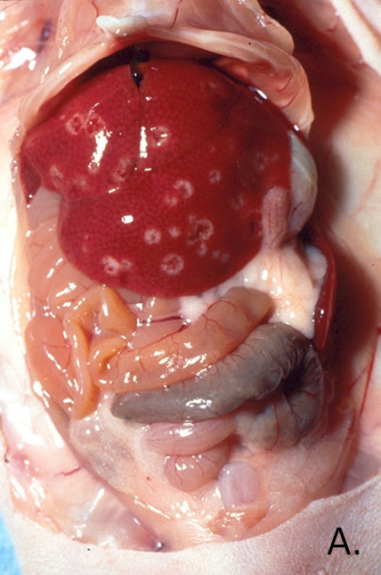
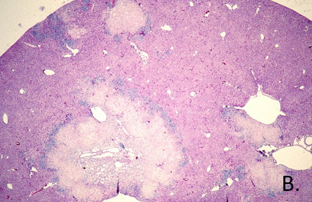
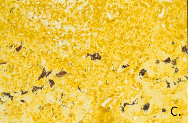
Pathology: In weanling or immunodeficient mice, serosal edema and hemorrhage in the ileocecocolic region of the gut and multiple yellowish-white foci of necrosis in the liver are prominent lesions (A.). Histological review of liver sections reveals coagulative to liquifactive necrosis with variable infiltrates of pyogranulomatous inflammatory cells (B.). Sections stained with silver help demonstrate large clumps of intracellular bacilli (C.) within hepatocytes bordering necrotic liver foci, and in the cytoplasm of the enterocytes in areas of granulomatous mucosal infiltrates.


Diagnosis: Diagnosis of C. piliforme is most commonly made by serology using IFA or MFI. PCR of lesions or feces can be used to diagnosis infection. Additionally, histopathology with demonstration of the bacillus in the tissues aided by the use of a silver stain can be used.