Etiology: Mouse adenoviruses are nonenveloped DNA viruses of the Mastadenovirus family. There are two strains of mouse adenovirus: mouse adenovirus type 1 (MAdV-1) and mouse adenovirus type 2 (MAdV-2).
Incidence: The incidence of infection is rare.
Transmission: MAdV-1 is transmitted through contact with infected urine. Mice that recover from acute infection shed MAdV-1 in the urine for prolonged periods (1-2 years) and viral DNA persists in brain, spleen and kidney for 1 year after experimental infection. Immunodeficient mice infected with MAdV-1 may also shed virus via fecal route.
MAdV-2 is shed in feces; the virus is transmitted via the fecal-oral route.
Clinical signs: Clinical signs depend on mouse age, mouse strain, virus species and viral dose and are found in mice experimentally infected with the virus. Neonatal mice develop fatal systemic disease with MAdV-1 infection. Adult mice of the susceptible strains (C57BL/6 and SJL/J) develop neurologic signs while the resistant BALB/c strain shows no evidence of disease except when experimentally inoculated with high dose of virus. No clinical signs are seen in mice naturally infected with MAdV-1 or MAdV-2.
Pathology: MAdV-1 infects cells of the macrophage lineage, renal tubular cells and vascular endothelial cells. In neonatal mice, necrosis from viral replication occurs in liver, spleen, kidney, brain and adrenal gland. Intranuclear inclusions are associated with foci of necrosis and can be found in most all tissues. In susceptible adult immunocompetent mice, hemorrhagic foci occur in the brain, especially the white matter, and are associated with viral induced damage to endothelial cells. Inclusions are uncommonly seen in brain lesions. Immunocompromised mice infected with MAdV-1 may display segmental hemorrhage in the mid small intestine; inclusions are not commonly observed.
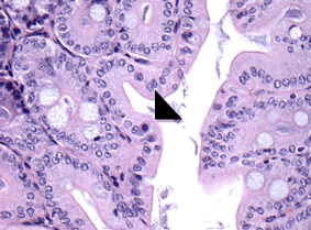
Intranuclear viral inclusions can be seen in infected enterocytes with MAdV-2 infection; viral-laden nuclei are often located in the apical portion of the cell (arrowhead).

Diagnosis: MAV infections can be confirmed by the detection of circulating antibodies. Virus can be amplified from infected tissues by PCR or cultivated in permissive cell lines. The enteric inclusions of MAdV-2 infection are pathognomonic for this agent.