Etiology: Syphacia muris is the most common pinworm found in rats, Syphacia obvelata is uncommonly seen.
Incidence: Incidence of infection is moderate.
Transmission: Transmission occurs via ingestion of ova.
Syphacia species adult females migrate from the cecum through the colon to the rectum, and deposit their eggs in a bolus on the perianal area. Eggs of Syphacia species are infective within 5-20 hours. The prepatent periods of Syphacia obvelata and Syphacia muris are 11-15 and 8-11 days, respectively.
Distribution: Adult Syphacia species are found in the cecum.
Clinical Signs: Clinical signs are not usually observed. Occasionally, heavy loads of pinworms may result in rectal prolapse or perianal irritation.
Diagnosis:
Antemortem:
Cellophane tape test of the perianal area should be used to detect ova of the Syphacia species.
Postmortem:
Place opened cecum and colon in a Petri dish containing saline. In a short amount of time, the pinworms will migrate out of the gut lumen into the saline. The pinworms can be detected with a dissecting microscope and speciated with use of light microscopy.
Diagnostic Morphology:
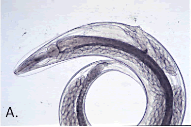
Syphacia muris: Round esophageal bulb. Small cervical alae (A.).
Female: 2.8 – 4 0 mm long. Vulva in anterior 1/4 of body.
Male: 1.2 – 1.3 mm long. Tail is long and pointed. 3 ventral mammelons: anterior mammelon is placed at the middle of the body, lengthwise.
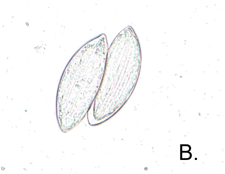
Ova: 72-82 x 25-36 µm. Thin-shelled, ellipsoidal, flattened on one side (B.).
Syphacia obvelata: Round esophageal bulb. Small cervical alae.
Female: 3.4 – 5 8 mm long. Vulva in anterior 1/6 of body.
Male: 1.1 – 1.5 mm long. Tail is long and pointed. 3 ventral mammelons. Center mammelon is placed at the middle of the body, lengthwise.
Ova: 118-151 x 33-55 µm. Thin-shelled, banana-shaped.

