Etiology:Clostridium difficileis aGram-positive, spore-forming, anaerobic bacilluswhich produces two toxins. Toxin A is an enterotoxin that binds to enterocyte receptors, causing fluid secretion and enterocyte damage associated with disease. Toxin B is a cytotoxin that causes cell death in vitro, but exact effects in vivo are unknown.
Incidence: Incidence of infection is moderate.
Transmission: C. difficileis transmitted by the fecal-oral route and overgrowth is precipitated by factors that disrupt gut flora such as diet changes and stress from shipping.
Clinical Signs: Death may be seen without observation of clinical disease. Anorexia may be seen several hours before the onset of diarrhea. Diarrhea is watery brown; guinea pigs are often moribund, dehydrated and hypothermic; death usually occurs 1 to 2 days after onset of diarrhea.
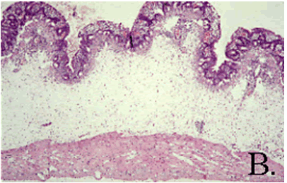
Pathology: Gross lesions include fluid cecal contents with serosal hemorrhages and edema on the cecum. Occasionally the ileum and proximal colon are affected. Microscopically, there is necrotic erosive or ulcerative typhlitis with swollen or vacuolated enterocytes, pseudomembrane formation; heterophilic infiltration, as well as submucosal edema and hemorrhage (B.). Large Gram-positive bacilli are present on the mucosal surface.
Diagnosis: Diagnosis of C. difficileis made by toxin analysis of feces, fecal PCR, culture (difficult) as well as evaluation of tissue lesions. Antigen detection ELISA can be used to detect toxins A and B.
