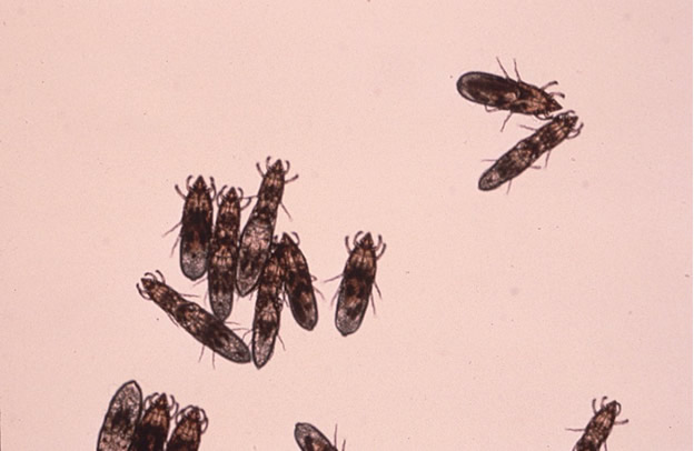
Etiology: Chirodiscoides caviae is the fur mite of guinea pigs (see picture).
Incidence: Mites are uncommon in guinea pigs.
Transmission: Transmission occurs by direct contact.
Distribution: Mites are most numerous over the rump and flanks.
Clinical Signs: Clinical signs are not usually observed. In heavy infestations, pruritus and alopecia may be evident.
Diagnosis:
Antemortem
- Pluck hairs and examine subgrossly (dissecting microscope) or microscopically for mites or eggs.
- Run cellophane tape against the grain of the fur, place on a slide and examine microscopically for mites or their eggs. This method is not very reliable for detection.
Postmortem
- Place pelage (fur) samples collected from the rump and perineum in a Petri dish. As the pelage cools, mites will migrate towards the tips of the hair shafts and be visible with a dissecting microscope.
- Place pelage samples on black construction paper. As the pelt cools, the mites will crawl away and be visible as brown specks on the black background.
Diagnostic Morphology: Elongated brown shield-shaped mite with 1st and 2nd pairs of legs adapted for clasping; posterior body is rounded in the female and blunted and triangular in the male.