Etiology: Encephalitozoon cuniculi is an obligate intracellular Gram-positive microsporidian parasite with a wide host range including rabbits, rodents, carnivores and man. Multiple strains of this parasite have been identified [1].
Incidence: The incidence of Encephalitozoon cuniculi infection is uncommon.
Transmission: There are two modes of transmission. The first is horizontal and occurs by the ingestion and possibly inhalation of the spores. Vertical transmission has also been reported in guinea pigs.
Clinical Signs: There are usually no clinical signs in acute or chronic infections.
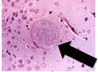
Diagnostic Morphology: Spores measure 2.5 x 1.5 µm (oval shape) with a thick wall (arrow).

Pathology: In acute cases the kidneys are swollen. Chronic lesions are more commonly seen and include multifocal, pinpoint, white, pitted areas on the surface of the kidneys. Histological examination of kidneys reveals granulomatous nephritis with spores in the tubular epithelia, interstitium or in tubular lumens. Examination of brain tissue reveals granulomatous meningoencephalitis with astrogliosis and perivascular lymphoid infiltrates. Organisms may be within glial cells or in granulomas.
Diagnosis: Diagnostic tests include serologic assays for detection of antibody to E. cuniculi and histopathologic demonstration of spores in infected tissues. PCR of kidney or brain tissue can also be used for diagnosis.
Public health Significance: E. cuniculi has been identified in immunosuppressed people.
1. Xiao, L., et al., Genotyping Encephalitozoon cuniculi by multilocus analyses of genes with repetitive sequences. J Clin Microbiol, 2001. 39(6): p. 2248-53.