Etiology: EDIM virus is a nonenveloped RNA virus of the rotavirus group A. Multiple strains of EDIM have been identified.
Incidence: The incidence of infection is low to moderate.
Transmission: Transmission occurs by fecal-oral, direct contact, and aerosol routes. Adult mice are unapparent viral carriers and shed the virus to their susceptible young.
Clinical Signs: Usually no clinical signs are noted in EDIM-infected mouse colonies. Neonatal mice with severe combined immunodeficiency (SCID mice) may be susceptible.
In experimentally-infected mice, a watery yellow diarrhea develops in 14-17 day old mice. Feces often dry on the perineum causing constipation and death. Surviving mice exhibit stunted growth.
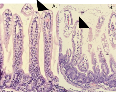
Pathology: Grossly, the intestines contain scant, yellow, gaseous contents. If a dried perianal fecal plug is present, the intestinal tract may be distended. Histopathologic features of the disease include vacuolar degeneration of infected enterocytes, typically at the villous tip (arrowhead, B.). Degenerative virus-induced vacuoles vary in size and are associated enterocyte nuclear pyknosis. Lesions induced with EDIM infection must be distinguished from the normal lipoprotein vacuoles seen in suckling mice, which are uniform and often contain a pink proteinaceous droplet, with unremarkable nuclei (arrowhead, A.).
Diagnosis: The clinical history and positive serological tests by MFI and IFA from dams of affected mice allow for confirmation of rotavirus infection. Additional diagnostic tests include PCR or commercially-available antigen capture ELISA of feces.