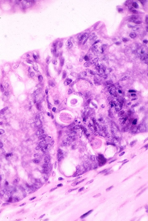
Etiology: Eimeria falciformis, Eimeria hansonorum, Eimeria ferrisi are coccidia of mice.
Incidence: Incidence of infection is rare. Young animals are primarily affected.
Transmission: Fecal-oral transmission via ingestion of sporulated oocysts. Oocysts require 1-3 days in an oxygenated atmosphere to sporulate.
Distribution:
E. falciformis: small intestine
E. ferrisi: cecum
E. hansonorum: small intestine
Clinical signs: Clinical signs range from none to enteritis which results in anorexia, diarrhea and sometimes death. Eimeria are often part of a multifactorial disease.
Relative pathogenicity:
E. falciformis > E. ferrisi, E. hansonorum
Diagnosis: Fecal flotation. Oocysts must be sporulated to spectate. Induce sporulation with time in a moist, oxygenated atmosphere or with 2.5% potassium dichromate solution.

Diagnostic morphology:
E. falciformis: 14-26 x 11-24 µm +/- polar granule; small steida body. No residuum or micropyle.
E. hansonorum: 16-18 µm, with a broad steida body.
E. ferrisi: 16-18 µm, with a small steida body.