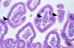
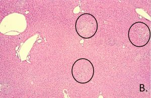
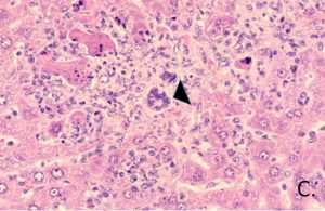
Histopathologic examination of the intestinal tract reveals transient syncytia (arrowhead, A.) of the mucosal epithelial cells in mice infected with enterotropic virus strains. Focal coagulative liver necrosis can be seen in mice infected with polytropic virus strains (B.). Hepatocellular syncytia are rarely present around necrotic foci in immunocompetent mice (arrowhead, C.). In immunodeficient mice, intestinal epithelial cells, hepatocytes and vascular endothelial cells undergo syncytial cell formation, necrosis and replacement with scarring in certain organs, especially liver.
Etiology: MHV is an enveloped RNA coronavirus. Approximately 25 strains or isolates have been described; several MHV strains display tropisms for different tissues.
Incidence: The incidence of enterotropic MHV strains is common while the polytropic strains are uncommon.
Transmission: Fecal-oral, direct contact, aerosols, and fomite transmission have been reported. Vertical transmission has been reported in experimental infections, but doesn’t appear to occur in spontaneous infections. MHV can also be transmitted through use of contaminated transplantable tumors or cell culture.
Clinical Signs: MHV is usually subclinical in immunocompetent mice. Disease expression is dependent on virus species and on host factors, including age, genotype, experimental status and immune function.
There are two major patterns of disease based on the tropism of the virus strain.
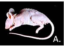
The respiratory (polytropic) pattern begins with virus replication in the nasal cavity and lungs with viremia and dissemination to other organs. Some strains may spread to the brain. Intestinal involvement is minimal. The enteric pattern infects the upper respiratory and intestinal tracts, with variable spread to other viscera. There may be overlap between these two disease patterns depending on which strain of MHV is involved. In suckling mice, watery diarrhea with mortality may occur with virulent enterotropic MHV infections. Immune deficient mice progressively lose weight and die (A.).
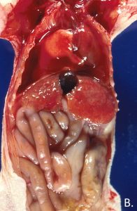
Pathology: Grossly, lesions of MHV infection in immunocompetent mice occur infrequently. Lesions, when present, may include gaseous distention of the intestinal tract in suckling mice, and multiple white liver foci in older mice. The liver may have a hobnail, nodular appearance (B.).
Diagnosis: Serology (MFI or IFA) of immunocompetent mice is the most useful diagnostic test for screening mouse colonies. PCR can be used to identify virus in feces. Histologic lesions are transient in immunocompetent mice; however histopathologic examination of target tissues may be helpful in identification of infection in immunodeficient mice.