Etiology: Syphacia obvelata and Aspiculuris tetraptera are nematodes commonly found in mice and other rodents.
Incidence: The incidence of pinworm infection is common.
Transmission: Infection occurs via ova ingestion. Eggs of Syphacia species are infective within 5-20 hours. Syphacia obvelata deposits eggs in the perianal region while Aspiculuris tetraptera releases eggs in the colon which then are passed in fecal pellets. The eggs of Syphacia obvelata are very light and have been shown to aerosolize, resulting in widespread exposure.
Distribution: Adult Syphacia species are found in the cecum. Aspiculuris tetraptera inhabits the proximal colon.
Clinical Signs: Clinical signs are not usually observed. It has been reported that heavy parasite loads may lead to rectal prolapse or perianal irritation.
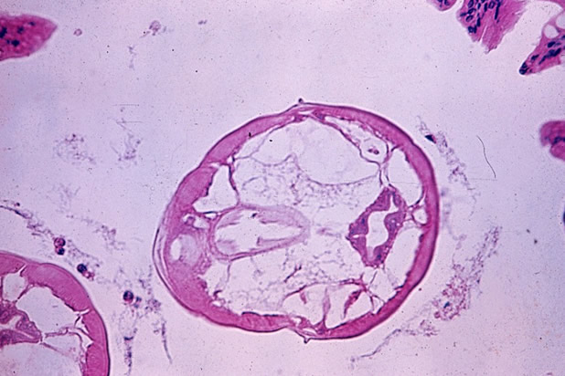
Pathology: On histology, cross-sections of pinworms can be identified in intestinal lumens (see photo, right).
Diagnosis: PCR of feces will detect pinworm infection. Direct exam of cecal or colonic contents will identify adults. Fecal flotation (for both pinworms) and tape test of the perianal region (for Syphacia only) will identify eggs.
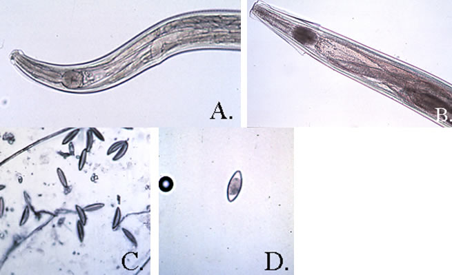
Diagnostic Morphology:
Syphacia obvelata: Round esophageal bulb. Small cervical alae (A).
Female: 3.4 – 5 8 mm long. Vulva in anterior 1/6 of body.
Male: 1.1 – 1.5 mm long. Tail is long and pointed. Three ventral mammelons; center mammelon is placed at the middle of the body lengthwise.
Ova: 118-153 x 33-55 µm, thin-shelled, banana-shaped (C).
Aspiculuris tetraptera: Oval esophageal bulb. Prominent cervical alae end abruptly at level of
esophageal bulb (B).
Female: 2.6 – 4.7 mm long. Vulva in the anterior 1/4 of body.
Male: 2.0 – 4.0 mm long. Tail is blunt and conical. No ventral mammelons.
Ova: 89-93 x 36-42 µm; morulated and football shaped (D).