Etiology: Pneumocystis murina is a fungal organism that historically had been recognized and misinterpreted to be a protozoan, hence the life stages are assigned protozoal descriptors such as trophozoites for the replicating stages and asci or cysts for the environmentally resistant forms. Pneumocystis murina is host specific.
Incidence: The incidence of infection with Pneumocystis murina is moderate.

Transmission: Cysts are horizontally transmitted by inhalation through direct contact with shedding mice. Immunocompetent mice are susceptible to infection, but clear the fungus and mount an immune response. Mice with defects in adaptive immune responses are more susceptible to persistent infection with development of pneumonia.
Clinical signs: In immunodeficient mice, signs may include dyspnea, weight loss, hunched posture; nude mice may have scaly skin.
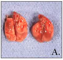
Pathology: Lungs do not deflate when the thorax is opened and may have patchy areas of consolidation (A.). Interstitial pneumonia with thickened alveolar septae from lymphocyte and macrophage infiltrates and proteinaceous, often foamy, material with activated macrophages in alveolar lumens are often appreciated (B.). Walls of cysts can be seen as 3-5 µm rings on sections stained with ammoniacal silver (PcAg of Grocott) or can be visualized using IHC (C.).
Diagnosis: Histologic recognition of cyst forms on sliver stained sections of lungs with typical pneumonia is commonly performed. Lung tissue can be subjected to Pneumocystis PCR. Lungs from immunocompetent mice are generally negative on Pneumocystis PCR due to the transient nature of their infection.