Etiology: Eimeria stiedae are eukaryotic, one-celled parasites.
Transmission: Transmission occurs by ingestion of sporulated oocysts.
Incidence: The incidence of infection is moderate to high.
Pathogenesis: Eimeria stiedae encysts in the duodenum, travels to the liver via the bloodstream or lymphatics, and invades epithelial cells of bile ducts to begin schizogeny.
Clinical Signs: Signs predominate in young rabbits and may include anorexia, debilitation, and pendulous abdomen with hepatomegaly noted on abdominal palpation. Mortality is low except in young rabbits. Diarrhea is uncommon.
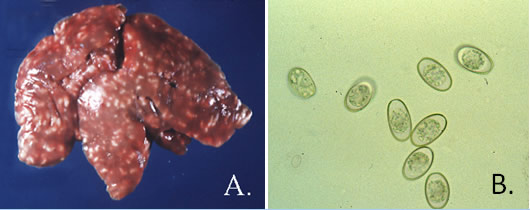
Pathology: An enlarged liver with multifocal, flat, yellow-white lesions containing yellow exudate (A.) and occasionally a distended gallbladder that contains bile may be seen at necropsy. The pathognomonic microscopic lesion is marked periportal fibrosis surrounding enlarged bile ducts lined with hyperplastic bile duct epithelium that harbors inflammatory cell infiltrates, and the presence of E. stiedae macrogametes, microgametocytes and oocysts.
Diagnosis: Multiple detection methods should be used. Radiographs can be used to visualize an enlarged liver or the presence of ascites. Diagnosis can be made by performing a fecal/bile flotation using supersaturated salt/sugar solutions, performing gall bladder scrapings or a direct smear of bile, and histologic examination of liver sections. Unsporulated oocysts are shown in (B).
