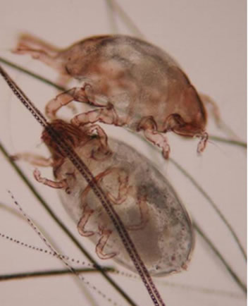
Etiology: L. gibbus is a small, nonburrowing mite. It is an obligate parasite, completing all stages of the life cycle on the host.
Incidence: The incidence of infection is uncommon. L. gibbus is less common than Cheyletiella parasitovorax.
Transmission: Transmission occurs by direct contact.
Distribution: Mites can be found over the entire body, but tend to concentrate in particular areas. L. gibbus is concentrated over the rump.
Clinical Signs: Clinical signs are not usually observed. Signs occasionally include a moist dermatitis which affects the back, groin, and ventral abdomen.
Diagnosis:
Antemortem
1. Pluck or brush hairs and examine subgrossly (dissecting microscope) or microscopically for mites or eggs.
2. Run cellophane tape against the grain of the fur, place on a slide and examine microscopically for mites or their eggs. This method is not very reliable for detection.
Postmortem
1. Place pelage (fur) samples collected in a Petri dish. As the pelage cools, mites will migrate towards the tips of the hair shafts and be visible with a dissecting microscope.
2. Place pelage samples on black construction paper. As the pelt cools, the mites will crawl away, and be visible as brown specks on the black background. *Be sure to ring the edges of the construction paper with double stick tape, so that the mites do not escape the area.
Diagnostic Morphology:Mites are brown, ovoid with short, ventrally directed legs. Males have a posterior clasping organ.
