Etiology: Fibroma virus is a DNA virus and a member of the leporipoxvirus group. It is closely related to myxoma virus.
Incidence: Rare in laboratory rabbits.
Transmission: Transmission is believed to occur from biting arthropods.
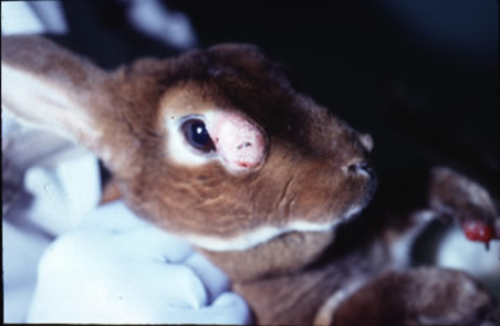
Clinical Signs: Discrete tumors occur on the legs or feet, on the muzzle, and around the eyes. Size depends on location of the tumor. The tumors are dermal and not attached to subcutaneous tissue. There is alopecia and rare skin ulceration over the tumors. The infected adult rabbit remains clinically normal otherwise. Tumors will typically regress after a period of about 6 months. Generalized disease and death may rarely be seen in young rabbits.

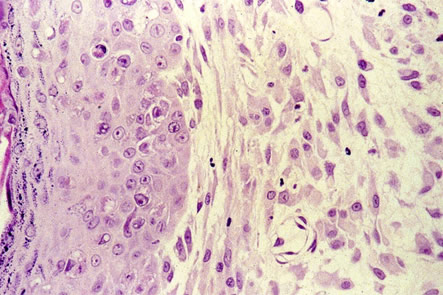
Pathology: Histologic appearance of fibromata may vary. Myxomatous fibromas contain large stellate mesenchymal cells in mucinous matrix (resembles myxomatosis). There may be fibroblast proliferation with minimal stroma. There may be intracytoplasmic eosinophilic inclusions in fibroblasts, stellate cells, and occasionally epidermis, as well as lymphoplasmacytic infiltrates around dermal vessels and at the base of the tumor. In generalized disease of young rabbits, fibromata present in many organs.

Diagnosis: Clinical signs and lesion morphology are the primary diagnostic tools. Virus isolation, serologic tests using IFA or serum neutralization using fibroma antibody can be used. PCR can also be used for diagnosis, but will not distinguish between fibroma virus and myxomavirus.