Etiology: Interstitial cell tumors are of Leydig cell origin.
Incidence: Common in aged F344 rats (up to 80%).
Clinical Signs: There are often no clinical signs.
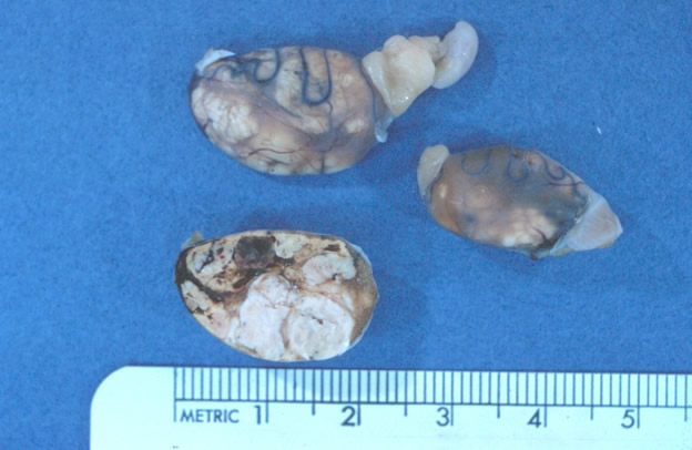
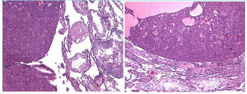
Pathology: Grossly, tumors are circumscribed, soft, lobulated, light yellow to hemorrhagic, single or multiple masses involving one or both testes. Histologically, tumors consist of 2 types of cells arranged in sheets. Cells are either polyhedral to elongated with granular to vacuolated cytoplasm or smaller with hyperchromatic nuclei and scanty cytoplasm.
Diagnosis: Diagnosis can be made upon necropsy and histopathologic examination of tissue.