Etiology: Entamoeba muris is a single-celled organism.
Incidence: The incidence of infection is common.
Transmission: Transmission is fecal-oral via ingestion of infective cysts.
Distribution: Amoebae are distributed in the cecum and colon.
Clinical signs: There are no clinical signs associated with infection. These protozoa may proliferate in diarrheic state, however, their role as contributors to disease is poorly understood.
Diagnosis:
Antemortem: Identification of cysts in feces is challenging, use of a sucrose gradient is necessary. Fecal PCR can be used.
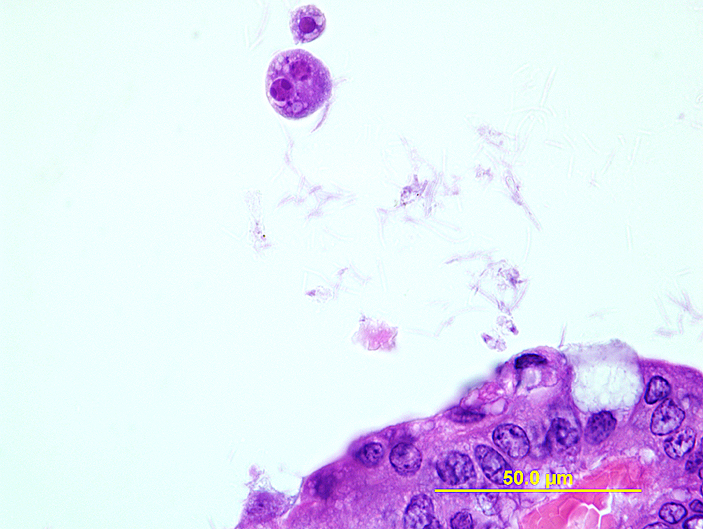
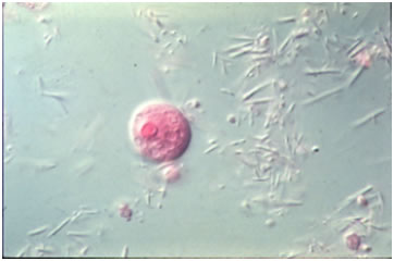
Postmortem: Wet mounts of cecal or colonic contents may reveal cyst forms, or globular trophozoite amoebas with slow extension of pseudopods.
Diagnostic morphology: 15-24 µm round cyst with 1-8 nuclei (nucleus number increases from 1 to 8 with maturity) and vacuoles. Immature cysts tend to have larger and more vacuoles than mature forms. Nuclei of both amoeba and cyst forms have eccentric karyosomes.
