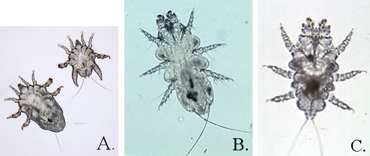
Etiology:Myocoptes musculinus (A.), Myobia musculi (B.), and Radfordia affinis (C.)
Incidence: The incidence of infection with fur mites is common.
Distribution: Myobia musculi and Radfordia affinis inhabit the hair shafts and skin surface of the intrascapular and dorsal cervical regions. Myocoptes musculinus is found on the hair shafts and skin surface over the flanks and rump in low numbers, and are distributed over the entire dorsum in high numbers. Fur mites feed on surface debris and do not invade the skin.
Transmission: Direct contact spreads the mite. Fur mites are usually host-specific.
Clinical Signs: Usually no clinical signs are observed. As an exception, heavy infestations have been associated with pruritus and poor looking hair coats. On genetically manipulated mice, mite infestations have been associated with pruritis, alopecia, epidermal excoriation and ulcerative dermatitis.
Diagnosis:
Antemortem
- PCR of skin swab or cage swab. This is the most reliable method for detection.
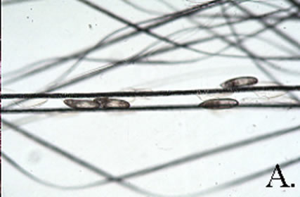
- Pluck hairs and examine subgrossly (dissecting microscope) or microscopically for mites or eggs (A.).
- Run cellophane tape against the grain of the fur, place on a slide and examine microscopically for mites or their eggs. This method is not very reliable for detection.
Postmortem
- Place pelage (fur) samples collected from the face and caput regions for Myobia musculi and Radfordia affinis, and from the rump or flank for Myocoptes musculinus in a Petri dish. As the pelage cools, mites will migrate towards the tips of the hair shafts and be visible with a dissecting microscope.
- Place pelage samples on black construction paper. As the pelt cools, the mites will crawl away and be visible as white specks on the black background.