Etiology: Lymphocytic Choriomeningitis Virus (LCMV) is an enveloped RNA arenavirus.
Incidence: Incidence of disease is rare in commercial breeding colonies.
Transmission: In utero or perinatal infections (within 1 day post-partum) produce a persistent, subclinical infection. If a mouse is infected after 24 hours of age, antibody production occurs. Virus is continually shed in urine, saliva and milk but antibody is difficult to detect due to low production and to binding with virus to cause circulating antibody-virus complexes. A major source of infection is transplanted tumors or cell lines.
Vertical (transovarian and/or transuterine) transmission is known to occur and is considered an efficient means of transmission to mice delivered by infected dams.
Clinical Signs: Two types of infections are known to occur. The persistent tolerant form results when the infection is acquired in utero or within a few days after birth. There is life-long viremia and shedding of virus. There is modest growth retardation, and at 7-10 months of age immune complex glomerulonephritis occurs and is associated with emaciation, ruffled fur, hunched posture, ascites and sometimes death.
The second, non-tolerant (acute) form occurs when mice are exposed after the first week of life. Viremia develops, but there is no shedding of virus. The outcome is either death within a few days or weeks, or recovery with elimination of the virus.
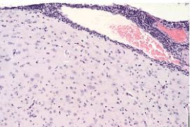
Pathology: Gross lesions vary from none to focal visceral necrosis and splenomegaly. Histological examination of the brain reveals lymphocytic infiltration of the meninges, choroid plexus, and of submeningeal and subchoroid perivascular spaces.

In tolerant mice, perivascular lymphocytic infiltrates of viscera and immune-complex glomerulonephritis may be observed. Please note that these lesions are also common in inbred strains of mice that are prone to development of autoimmune disease.
Diagnosis: Serologic testing using MFI and IFA help screen mouse colonies for the presence of infection. PCR of kidney or brain from infected mice or of biological samples will identify the virus. Virus isolation can be frustrating since LCMV does not induce a cytopathic effect. Bioassays include suckling mouse inoculation (footpad injection with infected neural tissue with observance of swelling within 5-9 days or cerebral injection with observance of neurological signs) and adult mouse inoculation (injection with whole blood and observation of clinical signs or antibody titers within 2 weeks).
Public Health Significance: The CDC reports human LCMV infections [3, 4].
3. Lymphocytic Choriomeningitis Virus From Pet Rodents. National Center For Infectious Disease Healthy Pets Healthy People 2012 July 28, 2010 [cited 2012 June 14, 2012]; Available from: http://www.cdc.gov/ncidod/dvrd/spb/mnpages/dispages/lcmv/owners.htm.
4. Barton, L.L., M.B. Mets, and C.L. Beauchamp, Lymphocytic choriomeningitis virus: emerging fetal teratogen. Am J Obstet Gynecol, 2002. 187(6): p. 1715-6.