Etiology: Sendai virus is an enveloped RNA paramyxovirus of the parainfluenza type 1 group.
Incidence: The incidence of infection is rare in laboratory animal facilities.
Transmission: Direct contact is the primary means of viral spread. The virus is not environmentally stable, but can be transmitted by fomites because of the quantities of virus excreted from infected mice.
Clinical Signs: Generally, no clinical signs are observed in mice in endemically infected colonies. In clinically apparent infections, signs are variable but may include chattering, mild respiratory distress, labored breathing, decreased fecundity in adults, deaths (possibly whole litters) in neonates and sucklings, and poor growth in weanling and young adult mice. In DBA/2 and immune deficient mice, the infection may be fatal. C57BL/6 mice are relatively resistant to clinical disease expression. This virus is immunosuppressive and may predispose to secondary bacterial infections.
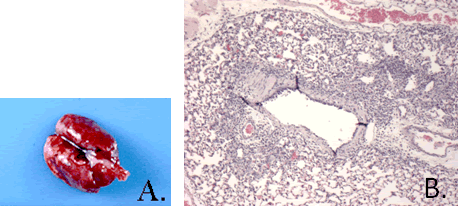
Pathology: Grossly, the lungs of affected mice may be mottled with red and tan foci in the parenchyma (A.). Exudate in the major airways may be seen. Histological examination reveals a characteristic interstitial pneumonia with perivascular and peribronchiolar lymphoid infiltrates and hyperplasia of alveolar macrophages (B.). Observation of squamous metaplasia of the bronchial epithelium is associated with the reparative stage of the infection. Lesions in resistant strains resolve quickly and eventually consist of loose peribronchiolar and perivascular lymphocyte cuffs.
Diagnosis: Commercially available MFI and IFA can be used to identify antibody titers in recovering mice. PCR of lung tissue can be used to diagnose Sendai virus in acute infections. Histologic lesions can be used to help diagnose infection in susceptible mice.